Muscle Dysfunction and Spine Health: How Advanced Diagnostics Are Changing the Way We Understand Back Pain
This post contains affiliate links. As an Amazon Associate, I earn from qualifying purchases at no extra cost to you.
Free resources — no credit card required for trial
🎧 Listen to health & wellness audiobooks free for 30 days
Start 30-Day Free Trial →
📚 Read unlimited health books free for 30 days
Try Kindle Unlimited Free →
If you’ve ever dealt with persistent back pain or a stiff, aching neck that just won’t budge, you’re far from alone. Millions of adults live with spinal discomfort every single day — and many of them never quite get to the bottom of what’s really causing it. We tend to focus on the obvious suspects: worn discs, compressed nerves, or misaligned vertebrae. But there’s a whole hidden world of muscle dysfunction in spinal pathologies that often goes undetected, quietly driving pain, instability, and frustration. The good news? Advances in diagnostic technology are giving doctors — and patients — a much clearer window into what’s actually happening deep inside the spine.
Why Your Spinal Muscles Deserve More Attention
Picture your spine as a tall, flexible mast on a sailing ship. What keeps it upright and steady, even as the waves crash? It’s not just the mast itself — it’s the intricate rigging of ropes and cables working in perfect tension around it. Your spinal muscles play exactly that role. In particular, the deep stabilising muscles like the multifidus and the transversus abdominis are the unsung heroes of your back. These aren’t the large, showy muscles you feel after a workout. They’re quieter, deeper, and arguably far more important for day-to-day spinal stability.
These muscles work around the clock to protect each individual segment of your spine, cushion your delicate nerves, and keep every movement smooth and controlled. When they’re functioning well, you barely notice them. When they’re not, everything can start to unravel. One muscle under-performing can cause others to overcompensate, creating a chain reaction of tension, fatigue, and pain that becomes increasingly difficult to break. Understanding this muscle dysfunction is absolutely central to addressing many forms of chronic spinal pain — which is why modern diagnostics are such a game-changer.
The Limits of Traditional Physical Examinations
When you visit a doctor or physiotherapist for back pain, the consultation usually starts with a hands-on assessment. They’ll look for visible signs of muscle wasting or imbalance, press along your spine to identify tender spots or areas of spasm, and test your strength, flexibility, and endurance. These assessments are genuinely valuable — they’re fast, accessible, and give a skilled clinician a useful starting picture of what’s going on.
But here’s the challenge: the deep stabilising muscles of the spine are buried beneath multiple layers of tissue. You simply cannot reliably assess the multifidus or transversus abdominis by pressing on someone’s back. It’s a bit like trying to diagnose a computer fault by looking at the keyboard. You might notice something is wrong, but you won’t necessarily know what is wrong or why. Traditional examinations also struggle to detect more subtle but highly significant changes inside the muscle tissue itself — things like fatty infiltration (where healthy muscle is gradually replaced by fat, a common finding in chronic back pain sufferers), microscopic inflammation, or abnormal patterns of muscle activation.
There’s also an element of subjectivity. Two experienced clinicians might interpret the same finding differently, which can lead to inconsistent diagnoses. For people with complex or persistent spinal conditions, this kind of uncertainty can mean months or even years of treatments that don’t quite hit the mark. That’s precisely why objective, technology-driven diagnostic tools have become so important in the modern management of spinal muscle dysfunction.
Advanced Imaging and EMG: A Closer Look at the Tools Transforming Spinal Diagnosis
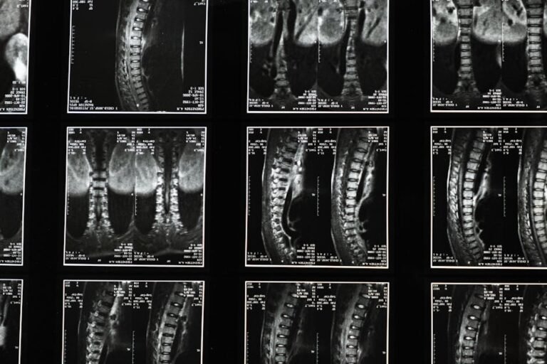
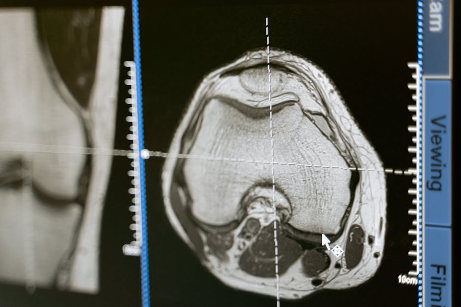
The diagnostic landscape for spine health has evolved dramatically. Three technologies in particular — MRI, ultrasound, and electromyography (EMG) — are helping clinicians identify specific patterns of muscle dysfunction in spinal pathologies with a precision that simply wasn’t possible before. Let’s break down what each one does and why it matters.
Magnetic Resonance Imaging (MRI) is best known for revealing disc herniations and nerve compression, but it’s also a powerful tool for examining muscle health. An MRI can show the internal architecture of spinal muscles in remarkable detail — including fatty infiltration, structural damage, and soft tissue inflammation. If a patient has been dealing with chronic low back pain for years, an MRI might reveal that a specific segment of their multifidus muscle has undergone significant degeneration, which helps explain why generic exercise programmes haven’t provided lasting relief. This structural information allows clinicians to target rehabilitation far more precisely.
Diagnostic ultrasound offers something that MRI cannot: real-time, dynamic imaging. Using sound waves, a trained practitioner can actually watch your muscles contracting and relaxing as you move. This means they can assess muscle thickness, activation timing, and coordination — all while you’re performing the movements that might normally trigger your pain. Even more excitingly, ultrasound can be used as a biofeedback tool during physiotherapy, letting you see your own deep muscles activating on a screen. For many patients, this visual feedback is transformative, helping them learn to engage muscles they didn’t even know they had.
Electromyography (EMG) takes a different approach entirely, measuring the electrical signals that travel between nerves and muscles. Every muscle contraction generates a tiny electrical impulse, and EMG captures these signals to reveal how well the nervous system is communicating with the muscle tissue. Is a muscle firing too weakly? Too strongly? At the wrong time relative to other muscles? EMG can answer all of these questions. It’s particularly useful for distinguishing between pain that’s primarily muscular in origin and pain driven by nerve dysfunction — a distinction that has major implications for how you should be treated.
Why Objective Data Makes Such a Difference for Your Back
The shift from subjective assessment to objective, measurable data represents a genuine leap forward in spinal care. When a clinician can point to specific findings — a measurable reduction in muscle cross-section, an abnormal EMG firing pattern, or visible atrophy on imaging — treatment decisions become far more confident and targeted. Instead of trying a range of interventions and hoping something sticks, the plan is built around what the evidence actually shows.
This matters enormously for patients. A diagnosis backed by objective data is easier to understand, easier to explain to other members of a care team, and easier to track over time. If you start a rehabilitation programme and return for a follow-up ultrasound three months later, your clinician can directly compare measurements and see whether your muscle is actually getting stronger and activating more efficiently. That kind of measurable progress is motivating, and it also allows for timely adjustments if something isn’t working as expected.
Objective diagnostics also help clinicians differentiate between the various spinal conditions that can cause similar symptoms. Disc herniation, spondylolisthesis (where one vertebra slips forward over another), degenerative disc disease, and facet joint problems can all produce overlapping patterns of pain — but the associated muscle dysfunction may look quite different under imaging or EMG. Identifying those distinct patterns is what allows for genuinely personalised care, rather than a one-size-fits-all approach.
What You Can Do: Practical Tips for Staying Ahead of Spinal Muscle Problems
Understanding that muscle dysfunction plays such a central role in spinal pain is actually empowering. It means there are concrete steps you can take — both in terms of how you engage with the healthcare system and how you look after yourself day to day. Here are some practical ways to be an active participant in your own spinal health:
- Be specific when describing your pain. Tell your doctor or physio exactly where it hurts, what type of sensation it is (sharp, dull, burning, aching), and what activities make it better or worse. The more detail you can provide, the easier it is to narrow down the likely source.
- Ask about advanced diagnostics. If your pain has been going on for more than a few weeks and hasn’t responded to initial treatment, it’s entirely reasonable to ask whether imaging, ultrasound assessment, or EMG could give you a clearer picture.
- Commit fully to rehabilitation. If you’re prescribed specific exercises — particularly core stabilisation work — take them seriously. Techniques like ultrasound biofeedback can help you learn to activate the deep muscles that are so crucial for spinal stability but so easy to neglect.
- Move regularly, but gently. Prolonged inactivity is one of the worst things for spinal muscles. Low-impact activities like walking, swimming, or yoga help maintain muscle tone and healthy blood flow to the tissues that support your spine.
- Work on your posture. Poor posture over time can contribute to muscle imbalances and dysfunction. Ergonomic seating, regular movement breaks if you work at a desk, and mindful posture during daily activities all add up.
- Maintain a healthy weight. Carrying excess weight puts additional load on spinal muscles and supporting structures, accelerating wear and contributing to the kind of fatty infiltration that advanced imaging can detect.
- Don’t hesitate to seek a second opinion. If a diagnosis doesn’t feel right or a treatment plan isn’t working, another qualified clinician’s perspective can be genuinely valuable.
The Future of Spine Health Is Personal — and It Starts Now
The way we understand and treat spinal problems is evolving rapidly. As these diagnostic tools become more widely available and more integrated into routine clinical practice, the days of vague diagnoses and trial-and-error treatment plans are gradually giving way to something far more precise and effective. The ability to identify specific patterns of muscle dysfunction in spinal pathologies — and to visualise them, measure them, and track them over time — means that both clinicians and patients can make far better-informed decisions.
For anyone living with chronic or recurring back pain, this is genuinely hopeful news. It means that if previous treatments haven’t worked, it may simply be because the full picture wasn’t visible yet. The right diagnostic tools, in the hands of a skilled clinician, can change that. And as a patient, your active involvement — in asking questions, committing to rehabilitation, and making supportive lifestyle choices — plays an equally important role in determining your outcomes.
Your spine carries you through every single day of your life. The muscles supporting it deserve every bit of attention, understanding, and care that modern medicine can offer — and now, more than ever, the tools exist to provide exactly that.
The Bottom Line: Muscle dysfunction in spinal pathologies is far more complex and influential than many people realise, and traditional physical examinations alone often can’t capture the full picture. Advanced diagnostic tools — including MRI, real-time ultrasound, and EMG — allow clinicians to detect structural changes, observe muscle activation patterns, and pinpoint nerve-muscle communication problems with a level of precision that’s transforming spinal care. If you’re struggling with persistent back or neck pain, understanding these tools can help you ask better questions, pursue more targeted treatment, and ultimately find more effective, lasting relief.
This is not medical advice. Consult your healthcare provider before starting any new health routine or using any product mentioned here.