MRI, Ultrasound, and EMG for Back Pain: How Advanced Spine Imaging Guides Personalized Treatment
This post contains affiliate links. As an Amazon Associate, I earn from qualifying purchases at no extra cost to you.
Free resources — no credit card required for trial
🎧 Listen to health & wellness audiobooks free for 30 days
Start 30-Day Free Trial →
📚 Read unlimited health books free for 30 days
Try Kindle Unlimited Free →
If you’ve ever been told you have back pain without getting a clear answer about why, you’re not alone. Living with spinal discomfort can feel like trying to solve a puzzle with half the pieces missing. Is it a strained muscle? A pinched nerve? A deeper structural issue? For years, doctors relied primarily on physical exams and patient-reported symptoms to make these calls — and while those tools still matter enormously, they don’t always tell the whole story. Today, advanced spine imaging techniques like MRI, ultrasound, and electromyography (EMG) are revolutionising the way spinal conditions are diagnosed and treated, making it possible to build truly personalised treatment plans based on what’s actually happening inside your body — not just educated guesses.
Why Your Deep Spinal Muscles Are So Difficult to Assess
Most people think of the spine as a column of bones, and while that’s technically true, the real magic — and the real vulnerability — lies in the muscles surrounding it. Your spine is supported by two types of muscle: the large, superficial muscles you can feel when you press on your back, and the smaller, deeper muscles that work quietly behind the scenes. These deep muscles, including the multifidus and transversus abdominis, are the unsung heroes of spinal stability. They activate during every movement you make, from sitting at your desk to lifting a bag of groceries, helping to protect and stabilise your vertebrae.
Here’s the problem: these deep muscles are incredibly hard to assess accurately using traditional methods. A physical examination can reveal visible asymmetry, surface-level tenderness, or general muscle spasm. But detecting whether your deep multifidus is functioning correctly? That’s a very different challenge. Subtle issues like muscle weakness, small fatty deposits within the muscle tissue, early scar formation, or minor swelling can easily slip through the cracks of a standard clinical assessment. And because physical exams are inherently subjective, two different clinicians might observe slightly different things — making it difficult to track your progress with precision over time.
This is exactly the gap that advanced diagnostic tools are designed to fill. By giving healthcare providers an objective, measurable look at what’s happening beneath the surface, these technologies transform the assessment of spinal muscle health from a guessing game into a science.
Seeing the Inside Story: How MRI and Ultrasound Reveal Spinal Health
When it comes to spine imaging, Magnetic Resonance Imaging (MRI) and ultrasonography are two of the most powerful tools available — and they each offer something unique.
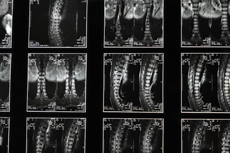
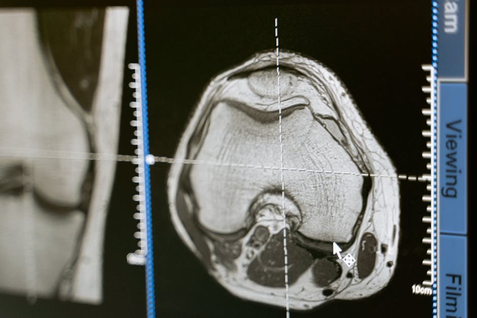
An MRI uses powerful magnets and radio waves to produce highly detailed cross-sectional images of your internal structures — muscles, ligaments, nerves, and bones alike. For spinal health in particular, MRI goes well beyond just spotting disc problems. It can reveal the actual size and shape of your deep spinal muscles, and — crucially — it can detect something called fatty infiltration. This is when healthy muscle tissue starts to be replaced by fat, which signals that the muscle isn’t functioning as it should. This isn’t simply a cosmetic concern; fatty infiltration in key spinal muscles is directly linked to chronic back pain and poor spinal stability. Knowing exactly which muscle segment is affected, and to what degree, gives physical therapists specific targets for rehabilitation — so instead of generic core exercises, you get a plan built around your actual weaknesses.
Ultrasound takes a different but equally valuable approach. Rather than producing a static image, it uses sound waves to create real-time, live visuals of your muscles as they move. This dynamic capability is incredibly useful for understanding how your muscles actually behave during activity — not just what they look like at rest. One of the most exciting applications is using ultrasound as a biofeedback tool during rehabilitation. Imagine watching your own deep core muscles on a screen as you try to engage them during an exercise. That immediate visual confirmation helps you learn to activate the right muscles in the right way, dramatically speeding up motor control retraining. For many people, this kind of real-time feedback makes the difference between months of frustrating progress and genuine, lasting improvement.
Listening to Your Muscles: What Electromyography (EMG) Can Tell You
While MRI and ultrasound give us a visual window into the spine, Electromyography — or EMG — takes a completely different approach. Instead of looking at muscles, it listens to them. Every time a muscle contracts, it generates tiny electrical signals. EMG technology measures these signals, offering a fascinating window into how well your nervous system and muscles are communicating with each other.
This matters more than you might think. A muscle can look perfectly healthy on an MRI and still fail to activate correctly when you move. EMG can detect these subtle discoordinations — moments when a muscle fires too late, too weakly, or not at all — that would be completely invisible on any imaging scan. This is what’s known as assessing neuromuscular control: the quality of communication between your brain and your muscles. Poor neuromuscular control is a significant contributor to spinal instability and chronic pain, and it’s something that can only be identified through electrophysiological testing.
One of the most clinically significant uses of EMG in spine health is its ability to help distinguish between muscle-based pain and neuropathic pain — pain that originates from nerve damage or irritation. This distinction is critical, because the treatment for each is fundamentally different. If a herniated disc is pressing on a nerve, for example, EMG can detect abnormal electrical activity in the muscles supplied by that nerve, helping to confirm nerve involvement and direct treatment accordingly. Similarly, EMG can identify specific patterns of muscle dysfunction associated with conditions like spondylolisthesis (where one vertebra slips over another) or chronic low back pain, giving your healthcare team the precise information they need to target the root cause of your symptoms.
From Guesswork to Precision: How These Tools Enable Personalised Treatment
Put together, MRI, ultrasound, and EMG elevate spinal assessment from a largely subjective art form to a far more objective and quantifiable science. Instead of relying solely on what you describe or what a clinician can observe, your healthcare team now has access to measurable, reproducible data about the actual state of your spinal muscles and nerves. This is a genuine game-changer — not just for diagnosis, but for the entire arc of your treatment and recovery.
The most meaningful benefit for you as a patient is the ability to receive a truly individualised treatment plan. Rather than being handed a standard sheet of generic core exercises, your programme can be designed around your specific findings. If MRI shows fatty infiltration in a particular muscle, your physical therapist can focus rehabilitation efforts on reactivating that specific muscle. If ultrasound reveals you’re not correctly engaging your deep stabilisers, biofeedback can guide you until you master the technique. If EMG confirms that nerve impingement is driving your muscle weakness, your doctor can pursue targeted interventions to address that nerve involvement rather than treating the symptoms alone.
This precision also means better monitoring of your progress over time. Because the data is objective and measurable, your healthcare team can track real changes in your muscle function, tissue health, and nerve activity — not just ask you how you’re feeling on a scale of one to ten. That kind of evidence-based progress tracking leads to smarter adjustments to your treatment plan and, ultimately, better long-term outcomes.
What You Can Do: Practical Tips for Navigating Spine Imaging and EMG
Understanding these advanced diagnostic tools is empowering. When you know what’s available and why it matters, you become a more informed and confident participant in your own care. Here are some practical steps you can take right now:
- Don’t accept vague answers: If you’ve been experiencing persistent back pain, spinal stiffness, or symptoms that haven’t responded to basic treatment, ask your doctor whether advanced spine imaging or electrophysiological testing might be appropriate for your situation.
- Ask what the results mean specifically for you: After any MRI, ultrasound, or EMG, ask your clinician to explain what the findings mean in plain language. Understanding your diagnosis helps you engage more meaningfully with your treatment plan.
- Embrace biofeedback during rehab: If your physical therapist uses real-time ultrasound during your sessions, lean into it. Watching your own muscles respond on screen is one of the most effective ways to retrain correct movement patterns.
- Ask about the “why” behind your exercises: If you’re given a rehabilitation programme, ask which specific findings are guiding each exercise. A well-informed patient is more likely to stay motivated and consistent.
- Track your progress actively: If you undergo baseline testing, ask whether follow-up assessments are planned so you can objectively measure improvement over time.
- Be honest about your symptoms: The more accurate and detailed the information you give your healthcare provider — including when pain occurs, what movements trigger it, and how long you’ve had it — the better equipped they are to order the right tests and interpret the results correctly.
- Seek out specialists when needed: If your current provider isn’t offering comprehensive diagnostic evaluations, consider asking for a referral to a specialist in musculoskeletal medicine, sports medicine, or a spine-focused physiotherapy practice.
The Bigger Picture: Why Precision Spine Care Matters for Your Quality of Life
Chronic back pain is one of the leading causes of disability worldwide, yet it remains poorly understood and frequently undertreated. Part of the reason is that back pain is not one condition — it’s dozens of different conditions that can look and feel remarkably similar on the surface. A pulled superficial muscle, a failing deep stabiliser, a compressed nerve, and a disc with fatty infiltration might all produce similar symptoms in day-to-day life, but they require very different treatments. Applying a one-size-fits-all approach to spinal care doesn’t just slow your recovery — it can actually make things worse by training the wrong muscles or ignoring the real underlying cause.
This is why the integration of advanced spine imaging and electrophysiology into routine spinal care is such an important step forward. It’s not about adding expensive tests for the sake of it. It’s about giving both you and your healthcare team the clearest possible picture of what’s actually happening — so that every decision made about your care is grounded in real data rather than assumptions. When treatment is targeted, specific, and evidence-based, you spend less time trying things that don’t work and more time making genuine, measurable progress toward a pain-free, active life.
The technology isn’t perfect, and not every clinic will have access to all of these tools. But knowing they exist — and knowing what questions to ask — puts you in a far stronger position as a patient. Advocating for a thorough diagnostic workup could be one of the most important steps you take on your road to recovery.
The Bottom Line: Advanced spine imaging tools like MRI, ultrasound, and EMG are transforming the way back pain and spinal conditions are diagnosed and treated. By offering objective, measurable insights into the structure and function of your deep spinal muscles and nerves, these technologies make it possible to move beyond generic treatment plans and into truly personalised care. Whether it’s detecting fatty infiltration in a deep stabilising muscle, using real-time ultrasound biofeedback to retrain motor control, or using EMG to distinguish nerve pain from muscle pain, these tools give both patients and clinicians a clearer, more accurate roadmap to recovery. If you’re struggling with persistent spinal symptoms, don’t be afraid to ask your healthcare provider about what advanced diagnostics might reveal — your back deserves that level of attention.
This is not medical advice. Consult your healthcare provider before starting any new health routine or using any product mentioned here.