Diagnostic Tools for Monitoring Treatment Efficacy: How MRI, Ultrasound, and EMG Are Changing Back Pain Care
This post contains affiliate links. As an Amazon Associate, I earn from qualifying purchases at no extra cost to you.
Free resources — no credit card required for trial
🎧 Listen to health & wellness audiobooks free for 30 days
Start 30-Day Free Trial →
📚 Read unlimited health books free for 30 days
Try Kindle Unlimited Free →
If you’ve ever felt like managing your back pain is a never-ending guessing game, you’re far from alone. One morning you wake up stiff and sore, the next afternoon a simple trip to the grocery store leaves you wincing. The frustrating truth is that back pain — particularly the persistent, chronic kind — is rarely simple. But here’s the encouraging news: the way doctors diagnose and monitor spinal problems has changed dramatically in recent years. Thanks to advanced diagnostic tools like MRI, ultrasound, and electromyography (EMG), healthcare providers can now peer deep inside your back, measure what’s really going on, and track whether your treatment is actually working. These aren’t just fancy gadgets — they’re transforming the path from pain to recovery.
Why Back Pain Is So Hard to Pin Down
Your spine is one of the most complex structures in your entire body. Think of it as the central pillar holding everything together, surrounded by an intricate network of muscles, nerves, ligaments, and discs. And it’s not just the big, obvious back muscles doing the heavy lifting. Deep beneath the surface lie small but critically important stabilising muscles — like the multifidus and transversus abdominis — that constantly make tiny adjustments to keep your spine safe and stable. When these deeper muscles aren’t working as they should, pain, instability, and injury can follow.
For decades, doctors have relied on physical examinations — looking at your posture, pressing on tender areas, testing your strength — to figure out what’s wrong. These methods are a good starting point, but they have real limitations. Those crucial deep stabilising muscles are simply too well hidden beneath layers of tissue to assess accurately by touch alone. It’s a bit like trying to diagnose a fault in your car engine by tapping the bonnet with your knuckles. You need to look inside.
Traditional physical assessments can also be subjective. Two clinicians examining the same patient might interpret their findings differently, leading to inconsistent diagnoses and treatment plans. They also struggle to detect subtle but important changes happening at the tissue level — like the early stages of fatty infiltration, where healthy muscle fibres start being replaced by fat, making those muscles weaker and less functional. For people living with chronic back pain, these nuances matter enormously. That’s where modern diagnostic tools come in.
Picturing the Problem: How MRI and Ultrasound Reveal What’s Really Going On
Two of the most powerful imaging tools available for spinal assessment are Magnetic Resonance Imaging (MRI) and ultrasonography. Both give clinicians a detailed window into what’s happening beneath the skin — but in different and complementary ways.
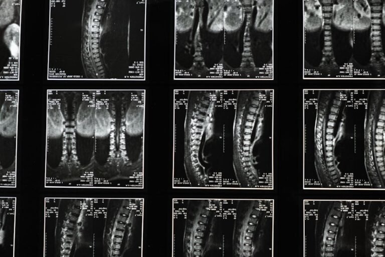
MRI uses powerful magnets and radio waves to produce highly detailed cross-sectional images of soft tissues, including muscles, ligaments, and nerves. Unlike X-rays, which are better suited to bones, MRI excels at visualising soft tissue. For spinal health, this means doctors can clearly see whether muscles are healthy, shrunken (atrophied), or showing signs of damage. Crucially, MRI can detect fatty infiltration in specific muscle groups — information that’s genuinely game-changing for treatment planning. If a key stabilising muscle is heavily infiltrated with fat, that tells your healthcare team that standard strengthening exercises alone probably won’t cut it. A more targeted rehabilitation approach — one focused on improving muscle quality, not just size — will likely be needed.
Ultrasound offers something MRI can’t: real-time, dynamic imaging. Using sound waves, ultrasound creates live images of your muscles as they move and contract. A physiotherapist can literally watch your deep abdominal or back muscles working (or failing to work) as you perform exercises right there in the clinic. This live feedback is incredibly valuable — not just for assessment, but as a teaching tool. Seeing your own muscles activating on a screen as you try to engage them is a powerful form of biofeedback. It helps you learn to activate the right muscles correctly, making your rehab exercises far more effective. Think of it as having a visual coach guiding your internal movements in real time.
Listening to Your Muscles: The Role of Electromyography (EMG) in Spine Health
While imaging shows us structure and movement, electromyography — more commonly known as EMG — takes a completely different approach. Muscles are controlled by electrical signals sent from your brain through your nervous system. EMG measures this electrical activity, giving clinicians a detailed read-out of how your muscles are actually functioning, and how well your brain and nerves are communicating with them.
For back pain patients, EMG can be genuinely revelatory. It can show which muscles are firing during movement, when they fire, and how strongly — information that can’t be gleaned from imaging alone. Someone might have muscles that look perfectly normal on an MRI, yet EMG reveals they’re activating too late, too weakly, or in the wrong sequence. These kinds of subtle coordination problems can place enormous strain on the spine over time, contributing to chronic pain and increasing injury risk.
EMG is also invaluable for distinguishing between muscular pain and neuropathic pain — pain that originates from nerve damage or irritation rather than the muscles themselves. This distinction is critically important because the two require very different treatment approaches. If a compressed nerve is causing muscle weakness, addressing the nerve problem takes priority. EMG gives clinicians the objective data they need to make that call confidently. Like ultrasound, EMG can also be used as a biofeedback tool, allowing you to see or hear your own muscle activity in real time and gradually learn to control it more effectively.
Monitoring Treatment Efficacy: Are You Actually Getting Better?
One of the most valuable — and often overlooked — applications of these diagnostic tools is tracking how well your treatment is working over time. It’s one thing to feel a bit better after a few weeks of physiotherapy; it’s another to have objective, measurable data showing that your muscles are genuinely recovering, that fatty infiltration has stabilised, or that your neuromuscular control is improving. That kind of evidence matters.
Monitoring treatment efficacy with tools like MRI, ultrasound, and EMG allows your healthcare team to make truly informed decisions about your care. If follow-up imaging shows that a previously underactive muscle is now contracting well, that’s a green light to progress your rehab programme. If results suggest a particular intervention isn’t producing the expected changes, your team can pivot early — before weeks or months are wasted on an approach that isn’t serving you. This is personalised medicine in action: treatment guided by concrete, measurable evidence rather than guesswork.
For people managing long-term or complex spinal conditions, this kind of objective monitoring can be genuinely empowering. Progress in back pain recovery isn’t always linear, and there will be days that feel discouraging. Having data that shows measurable improvement — even when it’s not yet fully felt — can provide crucial motivation and reassurance to keep going.
What You Can Do: Practical Tips for Getting Better Diagnostic Support
Understanding these tools puts you in a stronger position to advocate for your own health. Here’s how to make the most of what modern spinal diagnostics has to offer:
- Don’t accept vague answers: If your pain persists and treatments aren’t delivering results, know that more detailed evaluation options exist. You don’t have to settle for “let’s wait and see.”
- Ask about MRI or ultrasound: If you have ongoing pain or functional limitations, discuss with your doctor whether imaging could provide a deeper look at your spinal muscle structure — including checking for issues like fatty infiltration or muscle atrophy.
- Enquire about EMG: If there’s any suspicion of nerve involvement, or if your muscles seem to be poorly coordinated during movement, ask whether EMG could help build a clearer picture of your neuromuscular function.
- Embrace biofeedback-based rehabilitation: If your assessment reveals specific problems like poor deep muscle activation, ask your physiotherapist about incorporating ultrasound or EMG biofeedback into your sessions. It can dramatically improve the precision — and results — of your rehab.
- Use these tools to track progress: Diagnostic imaging and EMG aren’t just for initial assessment. Ask whether follow-up tests could be used to objectively measure how your treatment is going and guide any necessary adjustments.
- Look for an integrated care team: The best outcomes in spine health typically come from a collaborative approach — where a GP, physiotherapist, and specialist work together, combining clinical findings with advanced diagnostic data to create a truly holistic plan.
- Keep a symptom diary: Alongside formal diagnostics, tracking your own symptoms, triggers, and responses to treatment can provide valuable context for your healthcare team and help them interpret your results more accurately.
The Bigger Picture: Moving Towards Personalised Spine Care
The shift towards using advanced diagnostic tools in spine health represents something genuinely exciting: the move away from one-size-fits-all treatment towards care that is truly tailored to you as an individual. Two people might present with very similar back pain symptoms, yet have completely different underlying causes — one driven by nerve impingement, another by deep muscle dysfunction, another by fatty infiltration in specific muscle segments. Without the objective data these tools provide, those differences are easy to miss.
Spinal muscle evaluation is evolving from a largely qualitative art — dependent on the experience and interpretation of the clinician — into a more quantitative science, grounded in measurable, reproducible data. That doesn’t make the human element any less important; skilled, empathetic clinicians remain at the heart of great healthcare. But it does mean their decisions can be better informed, more precise, and more reliably effective.
For you as a patient, this evolution means a clearer path through the often-bewildering landscape of back pain treatment. It means fewer months spent on interventions that aren’t working. It means rehabilitation plans built around your specific weaknesses rather than generic protocols. And it means a greater sense of confidence and control over your own recovery — which, as any chronic pain sufferer knows, is worth an enormous amount.
The Bottom Line: If you’re dealing with persistent back pain and feel like you’re not getting the answers — or results — you need, it may be time to explore what advanced diagnostic tools can offer. MRI and ultrasound imaging, combined with EMG assessment, can reveal the real story behind your spinal symptoms, identify exactly where things are going wrong, and — crucially — help your healthcare team monitor whether your treatment is actually making a difference. You deserve care that’s based on evidence, not guesswork. Don’t be afraid to ask questions, advocate for thorough assessment, and push for a treatment plan that’s built around your body and your needs.
This is not medical advice. Consult your healthcare provider before starting any new health routine or using any product mentioned here.