Electromyography (EMG) and Your Spine Health: How Modern Diagnostics Reveal What’s Really Causing Your Pain
This post contains affiliate links. As an Amazon Associate, I earn from qualifying purchases at no extra cost to you.
Free resources — no credit card required for trial
🎧 Listen to health & wellness audiobooks free for 30 days
Start 30-Day Free Trial →
🛒 Recommended Products
As an Amazon Associate we earn from qualifying purchases at no extra cost to you.
Omron 5 Series Upper Arm Blood Pressure Monitor — 2-User 100-Reading Memory Wide-Range Cuf
$44.99
OMRON 7 Series Wireless Wrist Blood Pressure Monitor — Clinically Validated with Connect A
$69.99
OMRON Complete 2-in-1 Blood Pressure Monitor with EKG — Upper Arm Clinically Validated
$99.99
OMRON Silver Upper Arm Blood Pressure Monitor — Clinically Validated with Connect App
$49.99
OMRON Iron Upper Arm Blood Pressure Monitor — Clinically Validated Doctor Recommended for
$39.99
📚 Read unlimited health books free for 30 days
Try Kindle Unlimited Free →
If you’ve been living with persistent back pain, unexplained muscle weakness, or that nagging feeling that something just isn’t right — yet every standard test comes back inconclusive — you’re not alone. Millions of people find themselves stuck in a frustrating loop of vague diagnoses and treatment plans that never quite hit the mark. The truth is, traditional physical assessments, while useful, can only tell us so much. But thanks to remarkable advances in diagnostic technology, particularly a procedure called electromyography (EMG), healthcare providers can now “listen” to the electrical signals your muscles produce and gain a far deeper, more objective understanding of what’s driving your discomfort. This post breaks down exactly what EMG is, why it matters for spine health, and how it could be the key to finally getting the answers you’ve been searching for.
Why Traditional Muscle Assessments Often Leave You Without Answers
When you walk into a doctor’s office with back pain or muscle problems, the initial assessment typically follows a familiar pattern. Your provider will look for obvious signs like postural asymmetry or muscle wasting, press on areas to check for tenderness or spasm, and run you through basic strength and movement tests. These methods are quick, accessible, and genuinely helpful as a starting point. But they have real limitations — especially when the culprit is hiding deep beneath the surface.
Consider your deep core muscles — structures like the multifidus and transversus abdominis. These aren’t the visible “six-pack” muscles most people think of. They sit deep within your torso, wrapping around your spine like a natural internal corset, providing crucial segmental stability. Because they’re buried beneath layers of other tissue, there’s simply no reliable way to assess their true function through touch or observation alone. A practitioner pressing on your lower back can’t meaningfully evaluate whether your multifidus is activating properly during movement — it’s just too deep to feel with any precision.
There’s also the issue of subjectivity. One examiner’s interpretation of what they feel or observe can differ significantly from another’s, leading to inconsistent findings and, ultimately, inconsistent treatment. Subtle but clinically important changes — like early fatty infiltration of muscle tissue (where healthy muscle is gradually replaced by fat), fluid retention, or altered muscle activation timing — are virtually invisible to the naked eye or even a skilled pair of hands. This diagnostic gap is one of the main reasons so many people with chronic back pain feel like they’re going in circles, never getting to the root cause of their problem.
The Diagnostic Revolution: MRI, Ultrasound, and EMG Explained
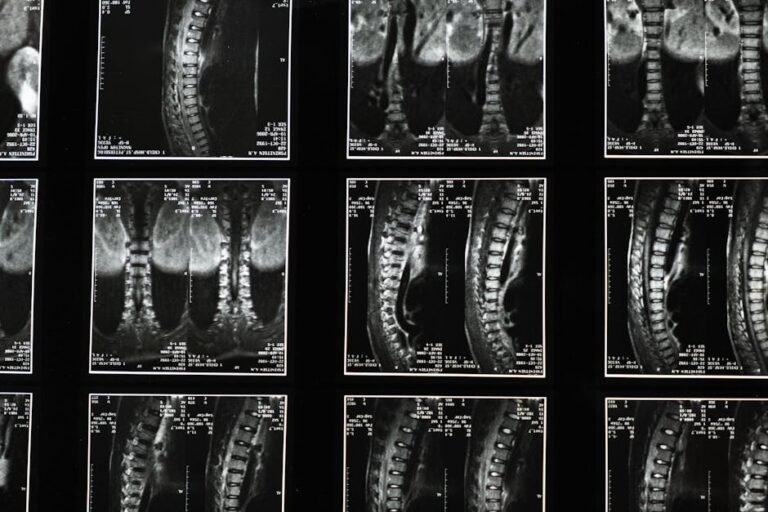
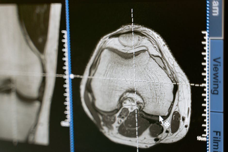
Thankfully, modern medicine has developed a powerful suite of objective diagnostic tools that go far beyond what any physical examination can reveal. Two of the most commonly used are Magnetic Resonance Imaging (MRI) and ultrasonography. MRI uses powerful magnets and radio waves to produce highly detailed images of soft tissues — it can reveal the architecture of your muscles, identify fatty infiltration, and assess tissue composition with a level of clarity that simply wasn’t possible a generation ago. Ultrasonography uses sound waves to generate real-time images, allowing clinicians to actually watch your muscles move and contract, measure their size and thickness, and even use what they see as biofeedback during rehabilitation.
But while MRI and ultrasound let doctors see your muscles, electromyography (EMG) lets them hear them. EMG is a specialised diagnostic procedure that measures the electrical activity your skeletal muscles produce. Every time your brain tells a muscle to contract, it sends an electrical signal down your spinal cord, through your nerves, and into the muscle fibres themselves. EMG captures those signals using small electrodes — either placed on the surface of your skin or, in more detailed assessments, inserted as fine needles directly into the muscle. The result is a precise, measurable record of how your nerves and muscles are communicating with each other.
Think of it like a sound engineer monitoring an orchestra. Each instrument — each muscle — should be playing its part at the right time, with the right intensity. EMG lets your healthcare provider hear whether every instrument is in tune, whether some are playing too softly, too loudly, or at entirely the wrong moment. It’s a level of insight that no physical examination, no matter how skilled, can replicate.
What Electromyography Actually Reveals About Your Spine and Muscles
The information an EMG provides is genuinely remarkable in its scope and specificity. For anyone dealing with spine-related conditions — chronic low back pain, disc herniation, spondylolisthesis, or nerve compression — these are the kinds of insights that can finally connect the dots between your symptoms and their source.
First and foremost, EMG can assess nerve function. If a nerve has been damaged or compressed — say, by a herniated disc pressing against a nerve root — the EMG will detect abnormalities in how that nerve is transmitting signals to the muscles it controls. This can help confirm or rule out nerve involvement as a driver of your symptoms. Second, EMG reveals how strongly and in what pattern your muscles are activating. Are they weak? Are they over-firing as a result of compensation? Are they switching on too late to properly protect a joint during movement? All of these details become measurable rather than guesswork.
Perhaps most valuably for spine health, EMG helps quantify what’s known as neuromuscular control — the sophisticated, largely automatic system by which your brain and nervous system coordinate muscle activity to stabilise and move your body. When this control system is disrupted, even subtly, it can lead to muscle imbalance, joint instability, and persistent pain that seems to resist treatment. EMG can pinpoint exactly where that disruption is occurring. It can also play a crucial role in distinguishing between pain that originates in the muscle itself and neuropathic pain caused by nerve damage — a distinction that is absolutely critical for choosing the right treatment approach.
From Diagnosis to Recovery: How Objective Data Changes Your Treatment Plan
Understanding the source of pain is only half the battle — the real value of electromyography and other advanced diagnostics lies in how dramatically they can improve the quality and specificity of your treatment. When a healthcare team can see objective, measurable data about your muscle and nerve function, they’re no longer working with educated guesses. They’re working with evidence.
This means your rehabilitation programme can be tailored specifically to you. If an EMG reveals that your multifidus isn’t activating properly on one side of your spine, your physiotherapist can design exercises specifically targeting that muscle and even use real-time biofeedback — from an ultrasound machine or surface EMG device — to help you actually feel and see that muscle working correctly during your sessions. This kind of precision rehabilitation is far more effective than a generic “core strengthening” programme that treats everyone the same way. If imaging reveals significant fatty infiltration in a particular muscle group, targeted strength and conditioning protocols can be prescribed to address that specific deficit.
Objective diagnostics also allow for meaningful monitoring of your progress over time. Rather than relying on subjective reports of whether you “feel better,” providers can track actual measurable improvements in muscle function, nerve conduction, and activation patterns. This not only keeps your treatment on track but also helps determine when it might be time to step up to more interventional approaches — or confirm that conservative management is working beautifully and should continue.
What You Can Do: Practical Tips for Getting the Most From Your Spine Health Assessment
Knowledge is genuinely empowering when it comes to your health. If you’re navigating spine pain, muscle weakness, or unexplained neurological symptoms, here are some practical steps you can take to advocate for yourself and make the most of modern diagnostic tools.
- Describe your symptoms in detail. Tell your healthcare provider not just where it hurts, but when the pain started, what makes it better or worse, whether it’s constant or intermittent, and how it affects your daily activities. The more specific you are, the more useful information your provider has to work with.
- Ask about your deep stabilising muscles. If you have chronic back pain, specifically ask whether the health and activation of your deep core and spinal stabilising muscles — like the multifidus and transversus abdominis — has been assessed. These are often overlooked in standard examinations.
- Inquire about advanced diagnostics. Don’t be afraid to ask your doctor whether EMG, MRI, or ultrasound might be appropriate for your situation. Understanding your neuromuscular control objectively could be the key to unlocking a more effective treatment plan.
- Ask “Why?” when you’re not getting answers. If your current treatment isn’t working or your diagnosis feels unclear, it’s entirely reasonable to ask for a more detailed explanation or to explore additional diagnostic avenues. Advocating for yourself is a healthy and important part of the healthcare process.
- Embrace biofeedback in rehabilitation. If your physiotherapist or rehabilitation specialist offers real-time ultrasound or surface EMG biofeedback during your sessions, engage with it actively. Being able to see or hear your muscles working correctly can dramatically accelerate the retraining process.
- Seek out reliable information. The more you understand how your body works — how your nervous system controls your muscles, why deep stability matters for your spine — the more effectively you can participate in your own recovery. Stick to reputable sources like peer-reviewed organisations, hospital websites, and qualified healthcare professionals.
- Be patient with the process. Objective diagnostics provide a clearer road map, but recovery from neuromuscular dysfunction or spine-related conditions still takes time and consistency. Trust the data, follow the tailored programme, and give your body the time it needs to heal.
The Bigger Picture: Why Objective Spine Health Assessment Matters
It’s easy to feel overwhelmed or discouraged when back pain or muscle dysfunction becomes a long-term part of your life. But it’s worth stepping back and appreciating just how far diagnostic medicine has come. Not long ago, providers were limited almost entirely to what they could see and feel in a clinical examination. Today, tools like electromyography allow us to measure the very electrical language your nervous system uses to communicate with your muscles — a level of insight that was once the stuff of science fiction.
For conditions involving the spine specifically, this matters enormously. The spine is a complex, dynamic structure that depends on an intricate network of muscles, nerves, and connective tissues all working in precise coordination. When that coordination breaks down — even subtly, in ways that are invisible to traditional assessment — the consequences can be significant: chronic pain, instability, compensatory movement patterns, and a quality of life that feels steadily diminished. EMG and complementary imaging tools like MRI and ultrasonography give healthcare providers the ability to identify those breakdowns with precision and address them directly.
The result is a healthcare experience that feels genuinely personal — not a one-size-fits-all prescription, but a treatment plan built around your specific muscles, your specific nerves, and your specific patterns of dysfunction. That’s not just better medicine. It’s medicine that respects the complexity of the human body and gives you a real, evidence-based path forward toward stronger, more resilient spine health and a better quality of life.
The Bottom Line: Electromyography (EMG) is a powerful diagnostic tool that measures the electrical activity of your muscles, revealing crucial information about nerve function, muscle activation patterns, and neuromuscular control that traditional physical assessments simply cannot provide. For anyone struggling with chronic back pain, spine conditions, or unexplained muscle weakness, EMG — alongside imaging tools like MRI and ultrasound — can transform the diagnostic process from subjective guesswork into precise, objective science. That clarity doesn’t just mean better answers; it means better, more targeted treatment, measurable progress, and a genuinely personalised path to recovery. If you’ve been searching for deeper answers about your spine health, asking your provider about advanced diagnostics could be one of the most important steps you take.
This is not medical advice. Consult your healthcare provider before starting any new health routine or using any product mentioned here.