What’s Really Going On in Your Back? How Advanced Spinal Muscle Imaging and EMG Can Reveal Hidden Problems
This post contains affiliate links. As an Amazon Associate, I earn from qualifying purchases at no extra cost to you.
Free resources — no credit card required for trial
🎧 Listen to health & wellness audiobooks free for 30 days
Start 30-Day Free Trial →
🛒 Recommended Products
As an Amazon Associate we earn from qualifying purchases at no extra cost to you.
Omron 5 Series Upper Arm Blood Pressure Monitor — 2-User 100-Reading Memory Wide-Range Cuf
$44.99
OMRON 7 Series Wireless Wrist Blood Pressure Monitor — Clinically Validated with Connect A
$69.99
OMRON Complete 2-in-1 Blood Pressure Monitor with EKG — Upper Arm Clinically Validated
$99.99
TRIDUCNA Shiatsu Neck Back and Shoulder Massager with Heat — Deep Tissue 3D Kneading Pillo
$49.99
Soothio Back Massager for Pain Relief Deep Tissue — Shiatsu 4D Motion with Heat and Car Ch
$89.99
📚 Read unlimited health books free for 30 days
Try Kindle Unlimited Free →
Have you ever woken up with a nagging back ache, tried stretching, maybe booked a massage, and still found yourself wondering — “What is actually going on in there?” You’re not alone. Millions of people live with back pain that seems to defy explanation, even after seeing a doctor. The truth is, the intricate world of spinal muscles, nerves, and bones is far more complex than any surface-level exam can fully reveal. That’s where modern spinal muscle imaging and electromyography (EMG) come in — powerful diagnostic tools that can peer deep inside your back and uncover hidden problems that traditional methods simply can’t detect. If you’ve been dealing with persistent back trouble and feel like you’re not getting the full picture, this article is for you.
Why Your Spinal Muscles Are So Hard to Assess From the Outside
Your spine is a genuine engineering marvel. It supports your entire body, allows you to bend, twist, lift, and move in countless directions — and it does all of this thanks to an elaborate team of muscles working around the clock. But here’s what many people don’t realise: the muscles doing the hardest work are the ones buried deepest, invisible to the naked eye and difficult to reach even with skilled hands.
We’re talking about deep stabilising muscles like the multifidus — a series of small but mighty muscles running along either side of your spine — and the transversus abdominis, a deep core muscle that essentially acts as a natural corset, wrapping around your midsection to keep everything stable. These muscles fire constantly during everyday movements, from sitting down at your desk to picking up a bag of groceries. When they’re working well, you barely notice them. When something goes wrong with them, the consequences can be significant.
Traditional clinical exams — where your doctor watches you move, checks your strength, feels for tenderness, and looks for muscle wasting — are genuinely valuable as a first step. But they have real limitations. They’re inherently subjective, meaning two different examiners might interpret the same findings differently. More importantly, they simply can’t detect subtle but meaningful changes happening inside the muscle tissue itself. Is a muscle slowly being replaced by fatty tissue? Is it activating at the wrong time? Is a nerve signal not getting through properly? These are questions that a standard physical exam often can’t answer — but modern technology can.
The Three Diagnostic Tools Transforming Spinal Muscle Assessment
Over the past few decades, advances in medical imaging and neurological testing have given healthcare providers extraordinary new ways to look inside the body. For spinal health specifically, three tools have emerged as particularly powerful: MRI, ultrasound, and EMG. Each offers a different kind of window into what’s happening with your spinal muscles and nerves.
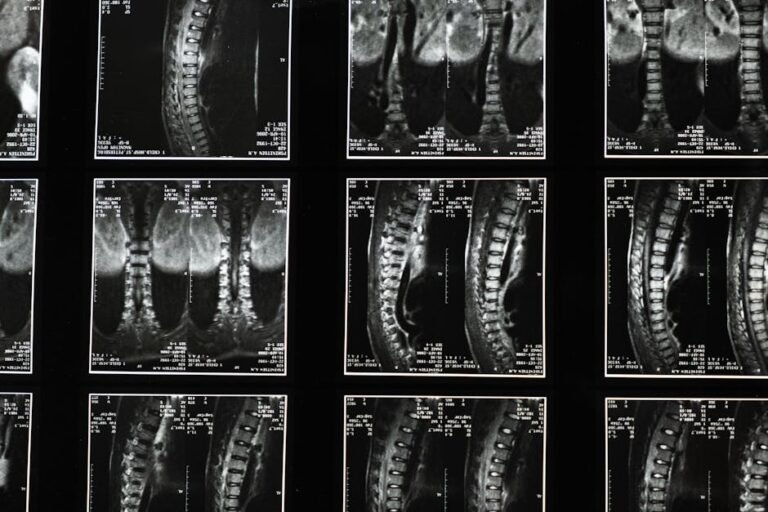
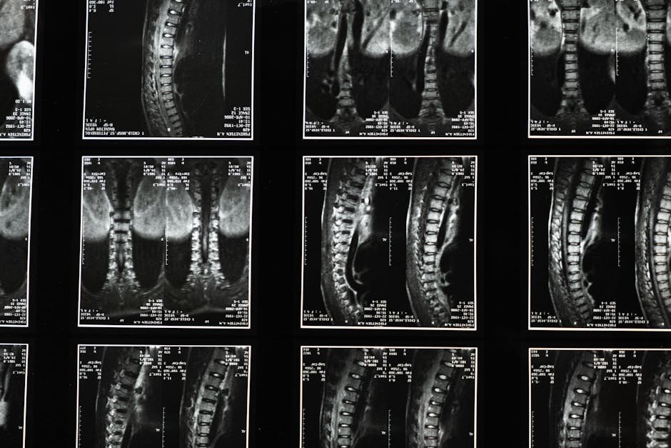
Magnetic Resonance Imaging (MRI) is probably the most familiar of the three. Think of it as taking incredibly detailed, high-resolution photographs of your body’s internal structures. When it comes to spinal muscles, MRI excels at showing their structure and tissue composition. Crucially, it can detect fatty infiltration — a process where healthy muscle tissue is gradually replaced by fat, which is a common finding in people with chronic back pain. This kind of information is completely invisible during a physical exam, yet it can fundamentally shape how your condition is understood and treated.
Ultrasonography (or diagnostic ultrasound) works differently — it uses sound waves to produce real-time images, almost like a live video feed of your muscles in motion. This makes it uniquely powerful for observing how muscles actually behave as you move and contract them. Clinicians can measure how much a muscle thickens when it activates, watch how it interacts with neighbouring tissues, and even gain insights into its overall health. Real-time ultrasound is also increasingly used as a biofeedback tool during physiotherapy, helping people literally see their muscles working as they practise exercises.
Electromyography (EMG) takes a completely different approach. Rather than producing images, EMG measures the electrical signals that travel from your nerves to your muscles — the signals that make your muscles contract in the first place. By recording and analysing these signals, EMG can reveal how well your nerves and muscles are communicating, whether certain muscles are overactive or underactive, and whether nerve damage might be contributing to your pain. This is especially useful for distinguishing between pain that originates in the muscle itself and pain that has a neurological root cause.
What Spinal Muscle Imaging and EMG Can Detect That You’d Otherwise Miss
The term “subclinical” might sound technical, but it simply refers to problems that are already present in your body before they’ve caused obvious symptoms or visible signs. This is one of the most important concepts in modern spinal care — because by the time a problem becomes obvious on a standard exam, significant changes may have already been happening beneath the surface for some time.
Consider fatty infiltration of the multifidus muscle, for example. Research has linked this change to chronic low back pain, yet a person might not realise it’s happening until it has progressed considerably. An MRI can catch this early, allowing treatment to be directed at the right target from the start. Similarly, EMG can reveal that a particular muscle isn’t firing properly — perhaps due to a nerve being compressed by a herniated disc — even when that muscle feels “normal” during a standard strength test.
These tools are also invaluable for pinpointing exactly which structures are involved in more complex spinal conditions. Whether someone has chronic low back pain, spondylolisthesis (where one vertebra slips over another), a disc herniation, or spinal stenosis, objective imaging and EMG data help paint a much clearer picture of what’s actually driving the problem. That clarity is the foundation of genuinely effective treatment.
How a More Precise Diagnosis Translates Into Better Treatment for You
Getting an accurate, objective diagnosis isn’t just satisfying from an intellectual standpoint — it has real, practical consequences for the quality of care you receive. When healthcare providers can see exactly what’s happening inside your back, they can design treatment plans that are targeted, personalised, and far more likely to work.
Take rehabilitation as an example. If an MRI reveals that a specific segment of your multifidus muscle has undergone significant fatty infiltration, your physiotherapist can design exercises that specifically target that area and encourage muscle recovery — rather than taking a generic “strengthen your core” approach. If an EMG shows that a particular muscle isn’t activating in the right sequence or at the right time, therapy can focus on re-educating that muscle’s timing and coordination, which is very different from simply building strength.
Real-time ultrasound and EMG biofeedback take this one step further by giving you immediate feedback during your exercises. Imagine trying to learn to play a musical instrument without ever being able to hear yourself play — that’s what retraining a deep spinal muscle can feel like without any feedback. With these tools, you can actually see or hear your muscle responding in real time, making it far easier to make corrections and build accurate movement patterns quickly.
Finally, these technologies allow genuine objective monitoring of your progress. Rather than relying purely on how you feel on a given day — which can fluctuate — measurable data from imaging and EMG can show whether your muscle structure, composition, and function are genuinely improving over time. This gives both you and your healthcare team solid evidence to work with, helping you stay motivated and allowing your treatment plan to be adjusted if needed.
What You Can Do: Practical Tips for Taking Charge of Your Spinal Health
Understanding that these advanced diagnostic options exist is the first step — but knowing how to navigate your healthcare journey with this knowledge is equally important. Here are some practical, actionable steps you can take:
- Advocate for yourself at appointments. If you’ve been dealing with persistent back pain that hasn’t responded well to standard treatments, it’s entirely reasonable to ask your doctor whether advanced imaging or EMG might be appropriate for your situation.
- Give your healthcare provider a detailed symptom history. Note where your pain is located, how intense it is, what makes it better or worse, how long it’s been going on, and what treatments you’ve already tried. The more information your provider has, the better equipped they are to recommend the right diagnostic approach.
- Don’t expect imaging for every ache. These tools are genuinely powerful, but they’re not necessary for every twinge or passing stiffness. Trust your doctor’s clinical judgement about when a deeper look is warranted — they’ll recommend it when symptoms are complex, persistent, or not responding to initial treatment.
- Be open to targeted, specialist therapies. If advanced diagnostics reveal specific muscle dysfunctions, embrace the opportunity to work with a physiotherapist who can use that information to design a highly personalised programme, potentially incorporating biofeedback techniques.
- Ask questions and seek to understand your results. If you undergo MRI, ultrasound, or EMG, ask your provider to explain what the findings mean in plain language and how they will directly influence your treatment plan. Understanding your diagnosis improves your motivation to follow through with treatment.
- Keep the bigger picture in mind. Objective diagnostics are a powerful part of spinal care, but they work best alongside a holistic approach. Regular appropriate exercise, good posture habits, ergonomic awareness at work, stress management, and a healthy weight all play important roles in supporting your spinal health long-term.
- Track your own progress. Keep a simple diary of your pain levels, functional abilities, and any changes you notice as you go through treatment. This personal record complements the objective data from clinical tools and helps your healthcare team see the full picture.
The Future of Spinal Muscle Diagnosis: Why This Matters More Than Ever
We’re living in an exciting era for spinal health care. Not so long ago, diagnosing what was truly happening inside the deep muscles of the back was largely a matter of educated guesswork — skilled guesswork, certainly, but guesswork nonetheless. Today, tools like MRI, diagnostic ultrasound, and EMG have transformed that process into something far more precise and evidence-based. The shift from qualitative observation to quantitative, measurable data is genuinely changing outcomes for people with back pain.
This matters particularly as our understanding of chronic back pain evolves. We now know that chronic pain is often driven by specific, identifiable changes in muscle structure and nerve function — changes that respond well to targeted treatment when they’re correctly identified. The old “rest and see if it improves” approach misses an important window of opportunity that objective diagnosis can open up.
As these technologies become more accessible and more widely used in clinical practice, more people will benefit from diagnoses that go beyond what the human hand and eye can detect. If you’ve ever felt that your back pain is more complicated than a standard consultation has been able to address, it may well be that the answers are there — they just need the right tools to find them.
The Bottom Line: Advanced spinal muscle imaging and EMG represent a genuine leap forward in our ability to understand and treat back problems. By revealing hidden changes in muscle tissue, composition, and nerve function that traditional exams can’t detect, these tools allow healthcare providers to diagnose more accurately, treat more precisely, and track progress more meaningfully. If you’re living with persistent or complex back pain, it’s worth having an informed conversation with your healthcare provider about whether these deeper diagnostic options might help you get the answers — and the relief — you’ve been looking for.
This is not medical advice. Consult your healthcare provider before starting any new health routine or using any product mentioned here.