The Paradigm Shift in Spinal Muscle Assessment: How Modern Imaging and EMG Are Changing Back Pain Care
This post contains affiliate links. As an Amazon Associate, I earn from qualifying purchases at no extra cost to you.
Free resources — no credit card required for trial
🎧 Listen to health & wellness audiobooks free for 30 days
Start 30-Day Free Trial →
🛒 Recommended Products
As an Amazon Associate we earn from qualifying purchases at no extra cost to you.
TruContour Lumbar Pillow for Sleeping — Adjustable Height Lower Back Support
$39.99
Branfit Shoulder and Back Brace Posture Corrector — Breathable Posture Trainer for Upper B
$24.99
Posture Corrector for Women and Men — Comfortable Effective Brace for Shoulder and Back Pa
$22.99
Bodywellness Posture Corrector for Men and Women — Adjustable Back Straightener with Clavi
$19.99
ComfiLife Lumbar Support Pillow for Sleeping — Memory Foam Triangle Wedge Pillow
$34.99
📚 Read unlimited health books free for 30 days
Try Kindle Unlimited Free →
Ever felt that nagging ache in your lower back and wondered why no one seems able to pinpoint exactly what’s going wrong? You’re not alone. Millions of people live with back pain that’s frustrating precisely because it’s so hard to understand — even for the doctors trying to help. The good news is that a genuine paradigm shift in spinal muscle assessment is underway, and it’s changing everything about how healthcare providers diagnose, understand, and treat the muscles that support your spine. Thanks to cutting-edge tools like MRI, ultrasonography, and electromyography (EMG), we now have an unprecedented window into what’s really happening deep inside your back. The result? More accurate diagnoses, more targeted treatments, and — most importantly — a real chance at lasting relief.
Why Spinal Muscles Have Always Been So Difficult to Assess
Think of your spine as the central pillar of your entire body. The muscles surrounding it are like the guy-wires of a suspension bridge — they keep everything stable, balanced, and capable of movement. While you can easily feel the larger, more superficial muscles across your back, the real workhorses for spinal stability sit much deeper. Muscles like the multifidus and transversus abdominis are nestled close to the vertebral column itself, providing fine-tuned, segment-by-segment support that acts almost like an internal shock-absorber system. These are the muscles that matter most for long-term spinal health — and they’re also the hardest ones to reach with traditional assessment techniques.
For decades, when you walked into a clinic with back pain, a practitioner would typically use a combination of visual inspection (looking for muscle imbalances or wasting), palpation (pressing gently to find tender spots or spasms), and manual muscle testing (pushing against your limbs to gauge strength). These approaches aren’t without value — they’re fast, accessible, and can reveal obvious problems. But trying to truly understand the deep stabilising muscles of your spine this way is a bit like trying to diagnose a car engine problem by kicking the tyres. You might notice something’s off, but you won’t really know what’s happening inside.
The limitations of these older methods are significant. It’s extremely difficult to accurately assess the strength or activation of a deep muscle like the multifidus through touch alone. Assessments can also vary depending on the examiner, meaning two different clinicians might reach different conclusions from the same patient. Perhaps most importantly, subtle but clinically meaningful changes — like the early onset of fatty infiltration within a muscle, mild swelling (oedema), or small shifts in how your muscles fire — can be completely invisible to traditional methods. Left undetected, these changes can contribute to chronic pain and long-term dysfunction. That’s why the shift towards objective, technology-driven spinal muscle assessment is such a significant development for anyone living with back pain.
A Closer Look Inside: The Imaging and EMG Tools Leading the Shift
Modern diagnostic technology is moving spinal muscle assessment from a largely subjective art into a more objective, data-driven science. Three powerful tools are at the heart of this transformation: Magnetic Resonance Imaging (MRI), ultrasonography, and electromyography (EMG). Together, they allow healthcare providers to directly visualise muscle architecture, measure how muscles activate, and assess the health of the nerves that control them — all with a level of detail and precision that simply wasn’t possible a generation ago.
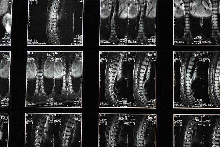
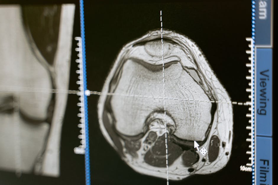
MRI: The Detailed Snapshot. If you’ve ever had an MRI scan, you know it involves lying in a large machine that uses powerful magnets and radio waves to create incredibly detailed images. For spinal muscle assessment, MRI is transformative because it doesn’t just show bones — it reveals the soft tissues surrounding them in extraordinary detail. Clinicians can use MRI to identify fatty infiltration within muscles (a common finding in people with chronic back pain that signals compromised muscle quality), detect oedema or swelling, and spot structural changes that would be completely invisible on a standard X-ray. Think of it as a high-resolution map of your muscle landscape, showing every change in tissue composition and structure.
Ultrasonography: The Real-Time Movie. While MRI gives a detailed still image, ultrasound provides something arguably even more useful for rehabilitation: a live, real-time view of your muscles as they move and contract. Using high-frequency sound waves, an ultrasound probe placed on your skin can show a muscle thickening as you engage it and thinning as you relax. This is incredibly valuable during rehabilitation — a therapist can actually show you on a screen whether you’re correctly activating a deep core muscle, providing immediate visual biofeedback. If you’ve ever struggled to “feel” whether you’re doing an exercise correctly, this kind of real-time visual confirmation is genuinely game-changing for learning proper muscle control.
EMG: The Muscle Whisperer. Muscles are powered by electrical signals sent from your brain through your nerves. Electromyography (EMG) measures this electrical activity, effectively “listening in” on the conversation happening between your nervous system and your muscles. Small electrodes placed on or near a muscle record these signals, revealing whether muscles are activating appropriately, whether certain muscles are working too hard or not hard enough, and whether there’s any abnormal electrical activity that might suggest nerve compression or damage. EMG is particularly valuable for distinguishing between pain coming from the muscle itself and pain with a neurological origin — like a pinched nerve — which is a crucial distinction when deciding on the right treatment approach.
From Data to Diagnosis: What These Tools Actually Reveal
The real power of imaging and EMG in spinal muscle assessment isn’t just about producing impressive-looking pictures or readings — it’s about what that information actually means for your care. These technologies allow clinicians to make critical distinctions that simply weren’t possible before. Is your back pain primarily a muscle issue, or is a compressed nerve driving your symptoms? Is a particular muscle weak and underactive, or is it actually working overtime to compensate for dysfunction elsewhere? Are your deep stabilising muscles engaging at all, or have they essentially “switched off” in response to pain or injury?
These aren’t trivial questions. The answers directly shape what kind of treatment will help you most. For example, if an MRI reveals significant fatty infiltration in a spinal muscle, the rehabilitation approach needs to focus on rebuilding genuine muscle quality and endurance — not just going through the motions of generic strengthening exercises. If ultrasound shows that a patient is completely unable to consciously activate their transversus abdominis (a deep core muscle critical for spinal stability), real-time biofeedback during therapy becomes an essential teaching tool. And if EMG identifies specific patterns of neuromuscular dysfunction linked to a disc herniation or chronic low back pain, the treatment plan can be precisely calibrated to address those exact issues.
This level of personalisation represents a genuinely new frontier in spinal care. Rather than a one-size-fits-all prescription of “rest and do some stretches,” clinicians equipped with objective data can design interventions that are tailored to what your specific muscles actually need. It’s the difference between guessing and knowing — and for people who have been struggling with back pain for months or years without finding real answers, that distinction matters enormously.
What This Means for You as a Patient
You might be thinking: “This all sounds impressive, but how does it actually affect my experience as someone with back pain?” The honest answer is that these advances matter most when your situation is complex, persistent, or not responding to standard treatments. If you’ve been dealing with chronic low back pain, have tried physiotherapy without much success, or have been told there’s “nothing to see” on a standard scan despite ongoing symptoms, the more detailed assessment tools described here could be precisely what’s needed to finally get to the bottom of your problem.
It’s also worth understanding that these tools are increasingly accessible. Ultrasound, in particular, is widely available in physiotherapy and sports medicine settings, and many clinicians now use it routinely as part of assessment and rehabilitation — not just in specialist centres. EMG is a well-established diagnostic procedure available through neurology and pain clinics. MRI, while more expensive, is a standard investigation for persistent or complex back pain in most healthcare systems.
Being an informed patient means knowing that these options exist and feeling empowered to ask about them. If your back pain has been dragging on without a clear explanation, it’s entirely reasonable to ask your healthcare provider whether advanced imaging or EMG might offer additional insight into what’s happening. A more precise diagnosis is almost always the first step towards a more effective treatment plan.
Practical Tips: What You Can Do to Support Your Spinal Health Right Now
Understanding the science behind spinal muscle assessment is genuinely empowering, but knowledge alone doesn’t resolve back pain. Here are some practical, actionable steps you can take today — alongside seeking appropriate professional care when you need it.
- Listen to your body. Don’t dismiss persistent back pain or ongoing stiffness as something you just have to live with. Minor aches can often resolve on their own, but chronic or worsening pain is a signal worth taking seriously.
- Stay active. Regular physical activity — including exercises that strengthen your core and back muscles — is one of the most well-supported strategies for spinal health. Even consistent daily walking can make a meaningful difference over time.
- Pay attention to posture. Be mindful of how you sit, stand, and lift throughout the day. Small ergonomic adjustments — like the position of your screen, the height of your chair, or how you pick things up from the floor — can reduce cumulative strain on your spinal muscles.
- Seek professional advice early. The sooner you consult a healthcare provider about persistent back pain, the sooner you can get on the right path. Early assessment often leads to better outcomes.
- Ask about your diagnostic options. If your back pain is complex or isn’t responding to initial treatment, ask your doctor or physiotherapist whether advanced imaging or EMG might be appropriate. Being an informed, engaged patient is one of your greatest assets.
- Commit to your treatment plan. Once a diagnosis is made and a plan is established, consistency is everything. Whether that means attending physiotherapy sessions, practising specific exercises at home, or making lifestyle adjustments, follow-through is what turns good advice into real results.
- Don’t neglect the basics. Quality sleep, stress management, and a balanced diet all contribute to muscle health and recovery. Spinal health doesn’t exist in isolation — it’s part of your overall wellbeing.
The journey towards better spinal muscle assessment is moving quickly, and the gap between what’s possible in leading research centres and what’s available in everyday clinical practice is narrowing fast. By staying informed and proactive about your own spinal health, you put yourself in the best possible position to benefit from these advances.
The Future of Spinal Muscle Assessment: A Brighter Outlook
It’s worth stepping back to appreciate just how significant this shift really is. For much of medical history, what happened deep inside the muscles of your spine was largely hidden from view. Assessment depended on a clinician’s experience, intuition, and the limited information available through touch and observation. That wasn’t a failure of medicine — it was simply the best available approach given the tools at hand. But it did mean that many people with genuine, significant spinal muscle dysfunction went undiagnosed or were given treatments that didn’t quite address the real problem.
The integration of MRI, ultrasonography, and EMG into spinal muscle assessment changes this picture fundamentally. These technologies allow healthcare providers to directly visualise muscle structure, observe function in real time, and measure neuromuscular activity with precision. They transform the evaluation of spinal health from an educated guess into an evidence-based investigation, with objective findings that can guide truly personalised treatment strategies. For patients, this means less time spent trying treatments that aren’t well-matched to the actual problem, and more time moving forward with approaches that are grounded in a genuine understanding of what’s happening in their specific spine.
None of this means traditional clinical skills are obsolete — far from it. The best care combines the human insight of an experienced clinician with the precision of modern diagnostic technology. But the addition of these tools to the assessment toolkit represents a genuine step forward for everyone dealing with back pain, from the person with a nagging ache that won’t resolve, to someone managing a more complex spinal condition. The paradigm has shifted — and it’s shifted in your favour.
The Bottom Line: Modern imaging and EMG technologies are genuinely transforming spinal muscle assessment, making it possible to see and measure what was once hidden from view. MRI reveals the structure and quality of your spinal muscles in precise detail, ultrasonography shows them working in real time, and EMG listens to the electrical signals that drive them. Together, these tools are moving spinal care from guesswork to evidence-based, personalised treatment — meaning better diagnoses and better outcomes for people living with back pain. If you’ve been struggling with persistent or unexplained back pain, it’s worth knowing these options exist and asking your healthcare provider whether they might be right for you.
This is not medical advice. Consult your healthcare provider before starting any new health routine or using any product mentioned here.